The Head And Neck Bones Anatomy And 3d Illustrations

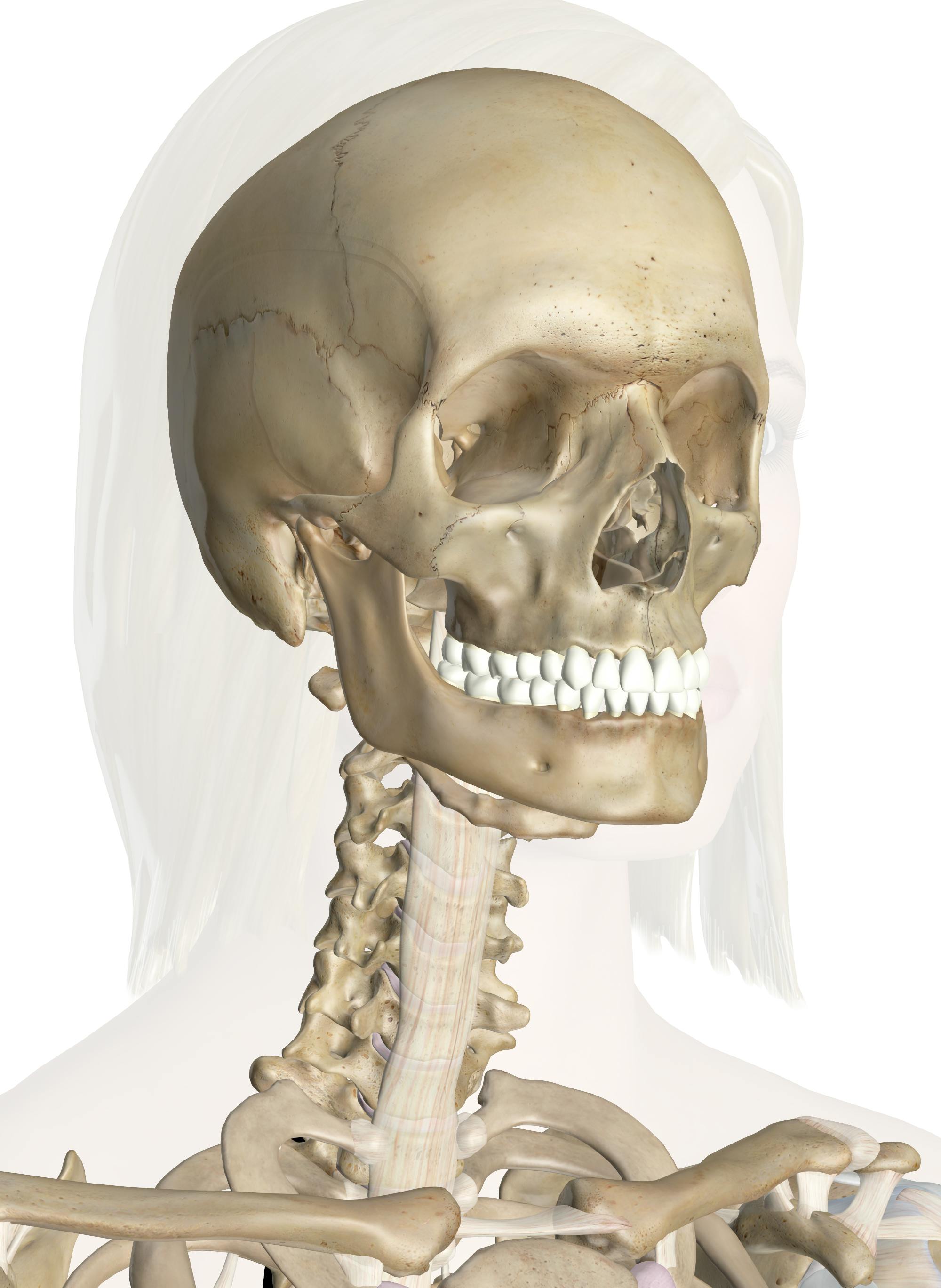
The Head And Neck Bones Anatomy And 3d Illustrations Explore the anatomy and vital role of the head and neck bones with innerbody's interactive 3d model. the bones of the head and neck play the vital role of supporting the brain, sensory organs, nerves, and blood vessels of the head and protecting these structures from mechanical damage. movements of these bones by the attached muscles of the. Orbit. bony orbit, extra ocular muscles, nerves of the orbit, lacrimal apparatus. anatomy.app unlocks the world of human anatomy. explore every muscle, bone, and organ! study interactive 3d models, articles, and quizzes that extend each other. an all in one platform for an efficient way to learn and understand anatomy.

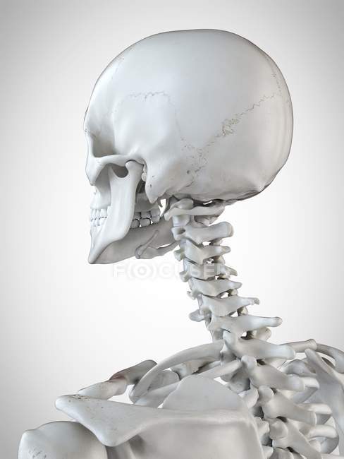
3d Rendered Illustration Of The Head And Neck In Human Skeleton Explore the anatomy and function of the cervical vertebrae with innerbody's interactive 3d model. the cervical vertebrae of the spine consist of seven bony rings that reside in the neck between the base of the skull and the thoracic vertebrae in the trunk. among the vertebrae of the spinal column, the cervical vertebrae are the thinnest and. Our team can also develop custom procedures demonstrating medical devices and disease states.**. the anatomy of the head and neck of the human body, including the bones, muscles, blood vessels, nerves, glands, nose, mouth, and throat. published 7 years ago. science & technology 3d models. blood. Composed of nasal cartilages and nasal bones, anterior to nasal cavity. arteries: facial, sphenopalatine, greater palatine, and ophthalmic arteries. nerves: olfactory nerve (cn i), ophthalmic nerve (cn v1), maxillary nerve (cn v2) eye. consists of ocular bulbs (eyeballs) with associated extraocular muscles located in orbits. The neck muscles, including the sternocleidomastoid and the trapezius, are responsible for the gross motor movement in the muscular system of the head and neck. they move the head in every direction, pulling the skull and jaw towards the shoulders, spine, and scapula. working in pairs on the left and right sides of the body, these muscles.

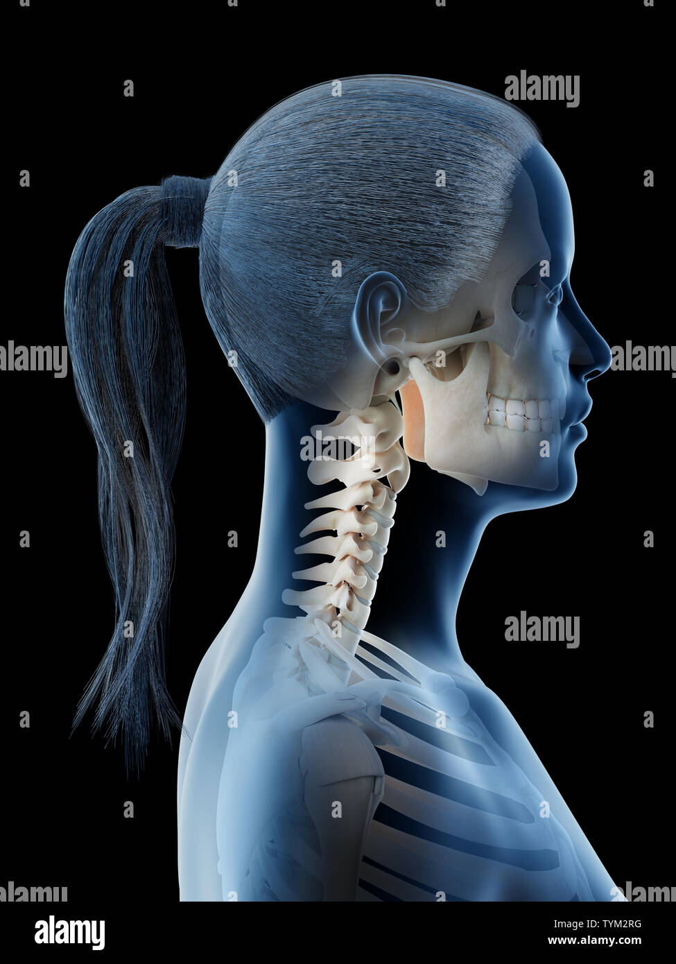
3d Rendered Illustration Of A Females Bones Of The Head And Neck Stock Composed of nasal cartilages and nasal bones, anterior to nasal cavity. arteries: facial, sphenopalatine, greater palatine, and ophthalmic arteries. nerves: olfactory nerve (cn i), ophthalmic nerve (cn v1), maxillary nerve (cn v2) eye. consists of ocular bulbs (eyeballs) with associated extraocular muscles located in orbits. The neck muscles, including the sternocleidomastoid and the trapezius, are responsible for the gross motor movement in the muscular system of the head and neck. they move the head in every direction, pulling the skull and jaw towards the shoulders, spine, and scapula. working in pairs on the left and right sides of the body, these muscles. Part of the teachme series. the medical information on this site is provided as an information resource only, and is not to be used or relied on for any diagnostic or treatment purposes. this information is intended for medical education, and does not create any doctor patient relationship, and should not be used as a substitute for. Figure 1 bones of cranium anatomy figure 2 cranium newborn : fontanelles figure 3 skull: anatomical illustrations figure 4 bony palate figure 5 cranial cavity , cranial sutures figure 6 internal surface of cranial base : superior aspect; vertical aspect figure 7 external surface of cranial base : inferior aspect figure 8 orbit.

Comments are closed.