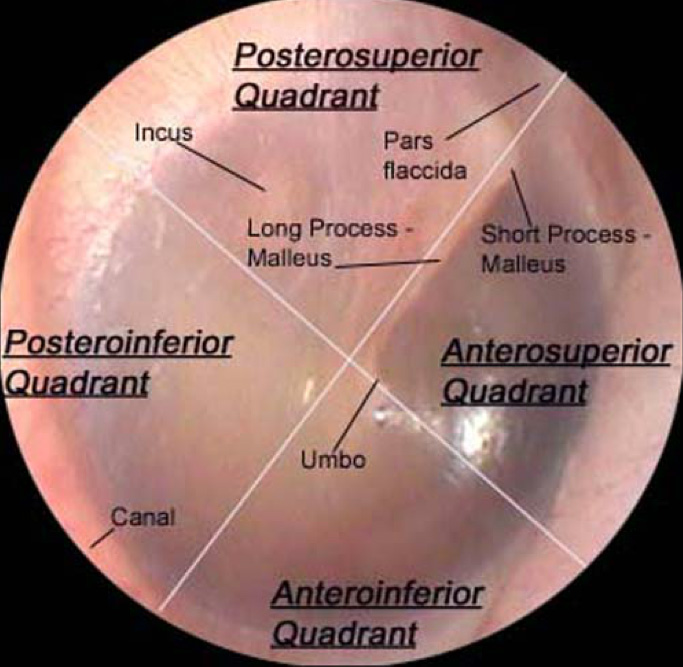
Normal Right Tympanic Membrane

Tympanic Membrane Anatomy вђ Department Of Pediatrics вђ Uwвђ Madison For a normal tympanic membrane, you should be able to observe*: lateral process of malleus; cone of light; pars tensa and pars flaccida *the cone of light can be used to orientate; it is located in the 5 o’clock position when viewing a normal right tympanic membrane and in the 7 o’clock position for a normal left tympanic membrane. Your tympanic membrane (eardrum) is a thin, circular layer of tissue that separates your outer ear from your middle ear. your eardrum plays an important role in hearing. it also protects your middle ear from dirt, bacteria and debris. contents overview function anatomy conditions and disorders care additional common questions.

Normal Tympanic Membrane Critical Care Practitioner The tympanic membrane, if visible, should be assessed for perforation, sclerosis, and retraction. the presence or absence of a normal light reflex should be noted. the attic area, immediately superior to the tympanic membrane, should be carefully inspected for signs of cholesteatoma. Tympanic membrane and middle ear. the tm separates the external ear from the middle ear. when inspecting the tm, the examiner takes note of color, bulging, perforation, and the presence or absence of normal landmarks. the cone of light, the handle of malleus, umbo, pars tensa, and pars flaccida make up the normal landmarks. The anatomy and function of normal hearing. sound is a vibration that enters the ear though the ear canal. this moves the tympanic membrane (“eardrum”) and the three small bones of hearing in the middle ear (malleus, incus, and stapes). the stapes vibrates as a piston against the fluid of the hearing postion of the inner ear (cochlea). The tympanic membrane (eardrum, myringa) is a thin, semitransparent, oval membrane, approximately 1 cm in diameter, that separates the external acoustic meatus from the tympanic cavity.[1][2] it is positioned at the lateral end of the external acoustic meatus and it is tilted medially from posteriorly to anteriorly and superiorly to inferiorly. therefore, the lateral surface of the tympanic.

Comments are closed.