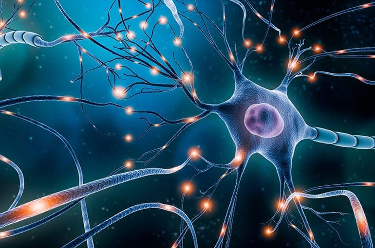
Neurons Firing Synapse

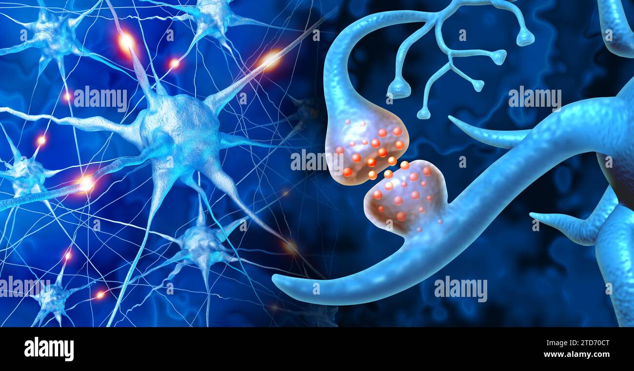
Neurons Firing Synapse It is best summarized by the mantra “neurons that fire together wire together.”. the idea is that neurons responding to the same stimulus connect preferentially to form “neuronal ensembles.”. these associations are mediated through synapses, the tiny connections through which neurons communicate and which can change through experience. In neuroscience, a synapse refers to the junction between two neurons, where information is transmitted from one neuron to another. it consists of the axon terminal of the sending (pre synaptic) neuron, the synaptic cleft (a small gap), and the dendrite or cell body of the receiving (post synaptic) neuron. neurotransmitters are released across.

Synapse Brain Neurology Human Brain Neurology And Cognitive Nerve The human brain is made up of approximately 86 billion neurons that “talk” to each other using a combination of electrical and chemical (electrochemical) signals. the places where neurons connect and communicate with each other are called synapses. each neuron has anywhere between a few to hundreds of thousands of synaptic connections, and. This model successfully predicted the firing rate modulation of individual output neurons across various network types with radically different response profiles (pre stdp, ie only, ee only, post. Synapse. diagram of a chemical synaptic connection. in the nervous system, a synapse[1] is a structure that permits a neuron (or nerve cell) to pass an electrical or chemical signal to another neuron or to the target effector cell. synapses are essential to the transmission of nervous impulses from one neuron to another, [2] playing a key role. Most brain neurons develop before birth, but the brain continues to mature long after that, with the neurons making and breaking an astonishing number of connections, called synapses. the neurons seen in this video were isolated from the cortex of a newborn mouse and grown in a dish where they were imaged every 30 minutes between days six and.

Comments are closed.